Details of DPV and References
DPV NO: 98 October 1972
Family: Alphaflexiviridae
Genus: Potexvirus
Species: Potato aucuba mosaic virus | Acronym: PAMV
Potato aucuba mosaic virus
B. Kassanis Rothamsted Experimental Station, Harpenden, Hertfordshire, England
D. A. Govier Rothamsted Experimental Station, Harpenden, Hertfordshire, England
Contents
- Introduction
- Main Diseases
- Geographical Distribution
- Host Range and Symptomatology
- Strains
- Transmission by Vectors
- Transmission through Seed
- Transmission by Grafting
- Transmission by Dodder
- Serology
- Nucleic Acid Hybridization
- Relationships
- Stability in Sap
- Purification
- Properties of Particles
- Particle Structure
- Particle Composition
- Properties of Infective Nucleic Acid
- Molecular Structure
- Genome Properties
- Satellite
- Relations with Cells and Tissues
- Ecology and Control
- Notes
- Acknowledgements
- Figures
- References
Introduction
- Described by
Quanjer (1921)
and
Clinch, Loughnane & Murphy (1936).
Selected synonyms
- Potato virus G (Rev. appl. Mycol. 16: 117)
- Potato calico virus (Rev. appl. Mycol. 14: 786)
- Potato virus F (= tuber blotch virus) (Rev. appl. Mycol. 16: 117)
- Potato calico virus (Rev. appl. Mycol. 14: 786)
-
An RNA-containing virus with filamentous particles about 580 nm long. It is transmitted by aphids in the non-persistent manner only when aided by certain ‘helper’ viruses, and is readily transmitted by mechanical inoculation.
Main Diseases
Causes yellow spotting or necrosis of potato leaves and necrosis in potato tubers, but is rare and consequently of little economic importance.
Geographical Distribution
World-wide but uncommon. The virus was prevalent in a few potato cultivars no longer grown commercially.
Host Range and Symptomatology
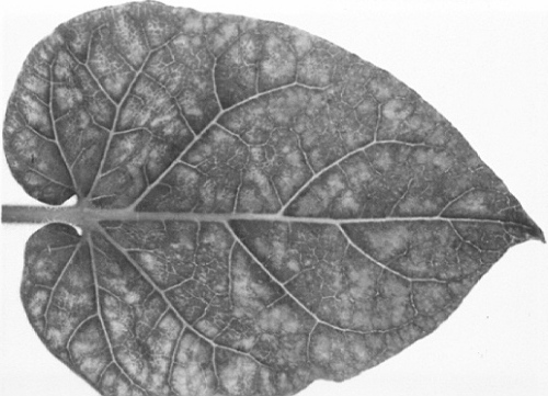
The type and intensity of the symptoms in potato plants depend on the strain of virus and the cultivar of potato. The two main types of symptom are: (1) bright yellow spots on the lower leaves, later coalescing to form large yellow or whitish patches (Fig. 1); (2) necrotic spots, often leading to systemic or top necrosis (Fig. 2) (Clinch, 1941). Some infected plants do not show foliage symptoms, especially during the second and subsequent years of infection and when growing under glass. During storage the tubers of many cultivars develop necrosis in the cortex and pith (Fig. 4) visible on the surface as irregularly shaped, brown patches or sunken dry brown areas (Fig. 3). Virulent virus strains may kill the tuber eyes.
-
Diagnostic species
- Capsicum annuum and C. frutescens (pepper). Necrotic local lesions
(Fig. 5)
followed by epinasty, systemic necrosis
(Fig. 6)
or severe mosaic; plants
infected when young are killed.
- Nicotiana glutinosa. Light green mottle with dark green vein-banding (Fig. 7).
- Lycopersicon esculentum (tomato). Some strains produce small round yellow spots on lower leaves.
- Nicotiana tabacum cvs. White Burley and Xanthi-nc, Datura stramonium and Petunia sp. may be infected systemically but are usually symptomless.
- Nicotiana glutinosa. Light green mottle with dark green vein-banding (Fig. 7).
-
Propagation species
- N. tabacum cv. Xanthi-nc is a good plant for maintaining the virus and a good source of virus for purification. Maximum virus concentration is found in inoculated leaves 10 days after inoculation using carborundum.
-
Assay species
- Capsicum annuum cv. Long Red and C. frutescens cv. Early Calwonder, and other cultivars.
Strains
Potato virus F, syn. tuber blotch virus (Clinch et al., 1936) is a minor variant causing mild leaf symptoms but typical tuber necrosis.
Transmission by Vectors
Transmitted by Myzus persicae in the non-persistent manner but only when aided by certain ‘helper’ viruses of the potato virus Y group, of which potato virus A and potato virus Y are the most efficient. Aphids transmit it from plants also infected with the ‘helper’ virus, or after feeding first on a source of ‘helper’ virus and then on a source of potato aucuba mosaic virus (Kassanis & Govier, 1971a; 1971b). In these conditions aphids can acquire the virus in 1 min. Starving aphids lose infectivity in a few hours.
Transmission through Seed
Not known; it is transmitted through the tubers produced by infected potato plants.
Transmission by Dodder
Not known.
Serology
The virus is strongly immunogenic and antisera with titres in excess of 1/10,000 are readily obtained. The virus gives flocculent precipitates in tube-precipitation tests.
Relationships
Strains of the virus are closely related to one another serologically and an avirulent strain protects potato plants from infection with a virulent strain. No serological relationship with other viruses has been demonstrated but potato aucuba mosaic virus resembles viruses of the potato virus X group morphologically.
Stability in Sap
In tobacco sap, the thermal inactivation point (10 min) of most strains is between 65 and 70°C, the dilution end-point between 10-5 and 10-6, and some infectivity remains after 1 to 2 months at 18 to 20°C in sap diluted 1/10, when inoculated using carborundum (Kollmer & Larson, 1960). The virus concentration is greater in the inoculated leaves of tobacco than in systemically infected ones, reaching a serological titre of 1/512 about 10 days after inoculation.
Purification
We find that the method described by Juo & Rich (1969) gives very aggregated preparations. The following method (D. A. Govier & B. Kassanis, unpublished) gives infective, largely unaggregated, preparations with yields of about 100 mg virus from 1 kg of leaves. Homogenize leaves in a cooled blender with 0.1 M ethylenediaminetetra-acetate pH 7.6 containing 0.01 M Na-diethyldithiocarbamate (DIECA), 2 ml/g tissue. Squeeze the sap through muslin, clarify by centrifuging for 30 min at 15,000 g, dialyse for 18-24 h against 10 volumes of 0.005 M DIECA, and reclarify. Shake the supernatant fluid for 5-10 min with 1/4 volume of chloroform, centrifuge, and subject the aqueous phase to 2 cycles of differential centrifugation (50 min at 100,000 g and 10 min at 15,000 g), resuspending the virus pellets in 0.01 M borate pH 7.5. Aggregation is largely prevented by leaving pellets covered with buffer overnight at 5°C before resuspending, by concentrating only slightly when resuspending the first high-speed pellets and, when a large weight of leaves is used, by infiltrating the leaves with extracting solution under vacuum before homogenizing them.
Properties of Particles
Sedimentation coefficient (s20,w) at infinite dilution: about 130 S.
Electrophoretic mobility: -0.7 x 10⁻⁵ cm2/sec-1/ volt-1 at pH 7 in 0.067 M phosphate buffer.
Absorbance at 260 nm (1 mg/ml, 1 cm light path): 2.6.
A260/A280: 1.1.
Particle Structure
Particles are filamentous, about 580 nm long and 11 nm wide (Fig. 8). They often aggregate end-to-end and side-to-side.
Particle Composition
RNA: About 5% of particle weight. Molar percentages of nucleotides: G25; A30; C24; U21 (J. M. Carpenter, unpublished).
Protein: Subunits have molecular weight of about 2.6 x 10⁴ (J. M. Carpenter, unpublished).
Relations with Cells and Tissues
Thin sections of infected tobacco leaves show virus particles in the cytoplasm, sometimes in dense aggregates. No other inclusions known (R. H. Turner, unpublished).
Notes
The yellowing of potato leaves resembles that caused by potato mop-top virus. Both viruses also cause necroses on the tuber surface, but those caused by potato aucuba mosaic virus are usually less clearly patterned than the raised necrotic rings caused by first-year infection with potato mop-top virus; however, sometimes potato aucuba mosaic virus causes necrotic rings. By contrast with potato aucuba mosaic virus, potato mop-top virus does not infect Capsicum annuum systemically, and has straight tubular particles about 20 nm in diameter (Harrison & Jones, 1970).
Figures

Systemically infected leaf of potato cv. Ninetyfold. (Courtesy Scottish Horticultural Research Institute.)

Potato cv. Majestic, systemically infected.

Infected potato tuber cv. Majestic.

Cut tuber of infected potato cv. Majestic.

Inoculated leaf of Capsicum annuum cv. Long Red.

Systemically infected Capsicum annuum cv. Long Red.
Systemically infected leaf of Nicotiana glutinosa.

Virus particles from a purified preparation. Bar represents 200 nm.
References list for DPV: Potato aucuba mosaic virus (98)
- Clinch, Scient. Proc. R. Dubl. Soc. N.S. 22: 435, 1941.
- Clinch, Loughnane & Murphy, Scient. Proc. R. Dubl. Soc. N.S. 21: 431, 1936.
- Harrison & Jones, Ann. appl. Biol. 65: 393, 1970:.
- Juo & Rich, Phytopathology 59: 1816, 1969.
- Kassanis & Govier, J. gen. Virol. 10: 99, 1971a.
- Kassanis & Govier, J. gen. Virol. 13: 221, 1971b.
- Kollmer & Larson, Res. Bull. agric. Exp. Stn Univ. Wis. 223, 40 pp., 1960.
- Quanjer, R. hort. Soc., Lond. Rep. int. Potato Conf. pp. 127-145, 1921.